Once the prospect of cancer looms in someone’s life, their first concern is to know exactly what the diagnosis is and what they need to do to start fighting it. That’s why new advancements in radiology that require fewer tests and provide more accurate data sooner are such an exciting development.
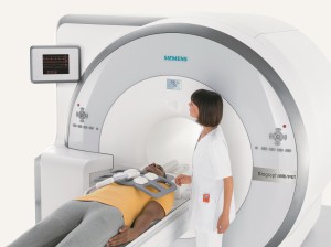
One of these is the MRI/PET (Magnetic Resonance Imaging/Positron Emission Tomography) scanner at Zwanger-Pesiri Radiology’s new office in Lynbrook. The FDA-approved Biograph mMR by Siemens has the ability to acquire both an MRI and a PET scan simultaneously, leading to better detection, characterization and treatment of oncologic diseases. According to Chief Executive Officer, Steven L. Mendelsohn, MD, there are nine of these units in U.S. research centers, but Lynbrook is the first private radiology facility to acquire it.
Typically cancer is diagnosed through the PET/CT (Computed Tomography) scan, which carries with it the dangers of radiation exposure. Some radiologists choose to use software techniques that combine a patient’s MRI scan with their PET scan—creating fused MRI/PET studies. But Dr. Mendelsohn said that due to motion and alignment issues with fusing two separate exams from two different scanners, diagnosing certain diseases can be problematic. He said the new MRI/PET is a diagnosis and staging machine that delivers better diagnostic information than the PET/CT and reduces radiation exposure equivalent to 100 chest X-rays.
“This dynamic test is for people who have a suspicion of a cancer or have a biopsy of other cancers to determine if they need surgery or chemotherapy,” said Dr. Mendelsohn. “It’s the best test we have for staging cancer based on size of the cancer, the local extension of the cancer and the spread to other organs. Without a question five to 10 years from now it will be the benchmark machine.”
Karen Mourtzikos, MD, director of molecular imaging, said the MRI/PET can also be used to see if a treatment is working or to detect a recurrence if a patient has been disease-free for an interval of time. The machine also improves detection for neurologic and cardiac diseases and for pediatrics.
“It’s hard to be sick and to have to navigate all this information [about cancer]. It’s like trying to take a sip from a fire hydrant. You have all this information blasting at you and it’s all very scary,” Dr. Mourtzikos said. “Now we have two tests in one and can say with greater certainty what we’re dealing with.”
The scans take between one to two hours and IV sedation is available. The results are given by radiologists the same day. Because Lynbrook is only 15 minutes from John F. Kennedy International Airport and a short ride from midtown Manhattan, Dr. Mendelsohn said he believes it will be accessed by many patients.
Unfortunately the MRI/PET test, which is costly, is not yet fully covered by insurance. It is recommended patients check with the facility to learn their options.
“Insurance companies did the exact same thing with MRI’s when they denied coverage in the late 80s and early 90s,” Dr. Mendelsohn said. “But people have cancer now. They cannot wait five to 10 years for it to be covered.”
What is covered in 2015 is an annual smokers’ screening test to detect lung cancer for adults aged 55 to 80 who have a 30-pack years (one pack a day for 30 years, two packs a day for 15 years, etc.) smoking history and currently smoke or have quit within the past 15 years. The low-dose computed tomography (LDCT) identifies the presence of lung cancer in an individual who does not demonstrate any symptoms. It is offered at 11 Zwanger-Pesiri offices.
There are advances in the detection of prostate cancer as well.
Byron Gaing, MD, whose specialty at Zwanger-Pesiri is body imaging, said that traditionally for those with suspected cancer urologists have been using ultrasound to guide a needle to do 12 random samplings
of the prostate.
“Certain spots of the prostate are prone to inaccurate samplings. But now that we have the MRI, when we see the lesions on the MRI we can fuse those images to the ultrasound machine,” Dr. Gaing said. “When radiologists do the biopsy, instead of aiming at a random area without actually seeing the target, they overlay a picture of the MRI so it’s more accurate.”
There are only a few of these machines in the area, including the Stony Brook office of Zwanger-Pesiri.
When it comes to screening for breast cancer, radiologists are recommending the ABUS or automated breast ultrasound system. Sharon Schlossberg, MD, whose specialty is body imaging and women’s imaging, said this screening tool shows an image of the entire breast. It is recommended for asymptomatic women with dense tissue.
“Mammography alone is not a sufficient screening tool if you have dense breasts or you test positive for the BRCA gene,” she said. “You can have this done on a yearly basis just like a mammogram.”
The ABUS is available in the Zwanger-Pesiri Plainview and Stony Brook offices.
For more information about updates in radiology visit www.zprad.com.